Rui Iun Cai,
Jie Wei ![]() ,
Feng Ling Jing,
Gui Zhen Zhao,
Yan Zhang
,
Feng Ling Jing,
Gui Zhen Zhao,
Yan Zhang
For correspondence:- Jie Wei Email: 119276645@qq.com
Received: 15 September 2016 Accepted: 8 January 2017 Published: 25 February 2017
Citation: Cai RI, Wei J, Jing FL, Zhao GZ, Zhang Y. Identification of metabolites of gardenin A in rat liver microsomes using ultra-high performance liquid chromatography coupled with linear ion-trap Orbitrap mass spectrometry. Trop J Pharm Res 2017; 16(2):421-427 doi: 10.4314/tjpr.v16i2.22
© 2017 The authors.
This is an Open Access article that uses a funding model which does not charge readers or their institutions for access and distributed under the terms of the Creative Commons Attribution License (http://creativecommons.org/licenses/by/4.0) and the Budapest Open Access Initiative (http://www.budapestopenaccessinitiative.org/read), which permit unrestricted use, distribution, and reproduction in any medium, provided the original work is properly credited..
Purpose: To identify the metabolites of gardenin A (GA) in rat liver microsomes (RLMs) using ultra-high performance liquid chromatography coupled with linear ion-trap Orbitrap mass spectrometry (UHPLC-LTQ-Orbitrap).
Methods: The sample was prepared by incubating GA (100 µg/mL) with RLMs (0.5 mg/mL) for 8 h. Then 5 µL of the sample was injected into UHPLC-LTQ- orbitrap mass spectrometer. The metabolites of GA were tentatively identified based on accurate mass measurements, fragmentation patterns, chromatographic retention times, and bibliography data.
Results: A total of 12 metabolites were detected and identified. Based on their structures, the main reactions in the metabolism of GA are de-methoxylation and de-methylation.
Conclusion: This is the first report on in vitro metabolites of GA. These results are considered very helpful for better comprehension of the metabolism of GA and its pharmacological effects.
Introduction
Murraya paniculata (L.) Jack, also called “qianlixiang in chinese” in China, belongs to the family Rutaceae. It is a very variable evergreen shrubby plant cultivated widely in gardens as an ornamental in many areas of China. The plant is officially listed in the Chinese Pharmacopoeia as a drug in Chinese Traditional medicine (TCM) [1]. Dried leaves or tender branches have been used all over China as a folk medicine due to the stimulant, astringent, antidysenteric, toothache remedy, and antidiarrheal [2-4]. Gardenin A (5-hydroxy-6, 7, 8, 3', 4', 5'-hexamethoxyflavone, GA), a hydroxylated polymethoxyflavonoid (OH-PMF), has been isolated from Murraya paniculata (L.) Jack [5-6]. Pharmacological studies indicate that it possesses variety of biological activities, such as anticancer and anti-inflammatory effects [7-9].
Research on drug metabolism research is very important in the early phases of drug discovery and development. It is also important for minimizing the adverse effect of pharmaceuticals, and for maximizing their therapeutic value [10,11]. A previous report [12] demonstrated that 26 metabolites of GA were observed in vivo; these might be responsible for the pharmacological actions of GA. However, it is hard to establish the reliability of this observation, due to difficulties involved in obtaining enough metabolites in vivo. Thus, it is important to obtain GA metabolites in vitro. To the best of our knowledge, metabolites of GA have not been investigated in vitro.
High performance liquid chromatography coupled with mass spectrometry has become a popular, and indeed the main method for the structural characterization of drug metabolites in vivo and in vitro due to its high efficiency, sensitivity, and selectivity. Among several different LC/MS platforms, ultra–high performance liquid chromatography high-resolution mass spectrometry (UHPLC-HRMS) [13-15] such as UHPLC-LTQ-Orbitrap is especially useful for the characterization of drug metabolites due to its higher and faster separation and resolution capacities.
The present study was carried out identify the metabolites of GA in rat liver microsomes (RLMs) with a view to providing better understanding of the metabolism of the drug and its pharmacological effects.
Methods
Chemicals and reagents
Gardenin A, 5-hydroxy-7, 3', 4', 5'-tetramethoxyflavone and 5-hydroxy-7, 3', 4'-trimethoxyflavone was purchased from Chengdu Biopurify Phytochemicals Co, Ltd (Sichuan, China). Acetonitrile, and methanol (HPLC grade) used were products of Fisher Scientific (Fisher, Fair Lawn, NJ, USA), and ultra-pure water used throughout the experiment was produced by a Milli-Q system (Millipore, Bedford, MA, USA). The 0.22 mm membranes used in the study were purchased from Waters Corporation (USA). Reduced form of nicotinamide adenine dinucleotide phosphate (NADPH) was supplied by Zhong Sheng Rui Tai Biotech (Beijing, China). RLMs were purchased from BD Biosciences (Bedford, MA, USA). All other reagents were of analytical grade.
Microsome incubation
Incubation conditions of microsome incubation experiment were established and controlled to provide a reproducible and linear rate of the metabolism in vitro. The incubation mixture in final volume of 1 mL contained GA (100 µg/mL), phosphate buffer (0.1 mol/L, pH 7.4), magnesium chloride (5 mM) and RLMs (0.5 g/mL). The incubation mixture was pre-incubated for 5 min in a water bath at 37 °C and reactions were initiated by addition of NADPH (1 mM). The reaction was terminated by adding 1 mL of ice-cold acetonitrile, vortexing and centrifuging at 15,000 rpm for 10 min at 4 °C. Aliquots of the supernatant were subjected to UHPLC-LTQ-Orbitrap MS to identify the metabolites. Blank samples were of the same composition as the test samples, but without GA. All samples were prepared in triplicate.
Instrumentation and chromatographic conditions
All UHPLC-MS analyses were carried out on LTQ/Orbitrap XL hybrid mass spectrometer (Thermo Electron, Bremen, Germany) equipped with an ESI source (Thermo Electron, Bremen, Germany). An Accela UHPLC system (ThermoFisher Scientific) was equipped with an autosampler, a vacuum de-gasser unit and a quaternary pump. Chromatographic separations were carried out on a BEH C18 column (2.1 × 50 mm, 1.7 μm) at room temperature and a flow rate of 0.2 mL/min. The mobile phase consisted of solvent A (water) and solvent C (acetonitrile). The percentage of solvent A was changed as follows: 0-2 min, 95 % A; 2-3 min, 95-80 % A; 3-20 min, 80 - 40 % A; 20-21 min, 40-20% A; 21-25 min, 20 % A; 25-26 min, 20-95 % A; 26-30 min, 95 % A; an aliquot of 5 μL of the supernatant was injected into UHPLC-LTQ-Orbitrap MS.
The optimized operating parameters in the positive ion mode were as follows: capillary voltage of 25 V, source voltage of 4.0 kV, capillary temperature of 350 °C, sheath gas flow rate of 40 (arbitrary units), auxiliary gas flow rate of 20 (arbitrary units), and tube lens of 110 V. Metabolites were detected by full-scan mass analysis from 100 to 800 m/z at a resolving power of 30,000 with data-dependent MS2 analysis triggered by the three most-abundant ions from the precursor list of predicted metabolites followed by MS2 analysis of the most−abundant product ions. Collision-induced dissociation (CID) was performed with an isolation width of 2 Da. The collision energy was set to 35 %.
Data processing
Thermo Xcaliber 2.1 workstation (Thermo Fisher Scientific) was used for data acquiring and processing. In order to obtain as many fragment ions as possible, the peaks detected with intensity over 50,000 were selected for identification. The chemical formulas of all parent and fragment ions of the selected peaks were calculated from the accurate mass using a formula predictor by setting the parameters as follows: C [0 - 35], H [0 - 50], O [0 - 15], S [0 - 1], N [0 - 3], and Ring Double Bond (RDB) equivalent value [0 - 15]. Other elements such as P and Br were not considered as they were rarely present in the complex matrix. The maximum mass errors between the measured and the calculated values were < 5 ppm. Blank RLMs samples were used as control to compare with the analyzed samples, and they were all processed under the same conditions.
Results
Fragmentation pathway of GA
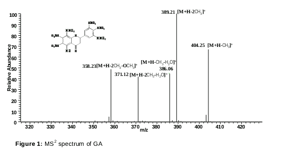
The parent ion showed a protonated ion [M+H]+ at m/z 419.13321 (-1.07 ppm, C21H23O9) in positive ion mode. Fragmentation of this parent ion at m/z 389, m/z 404, m/z 358, m/z 371, and m/z 386 by the loss of the moiety 30 (2CH3。), 15 (CH3。), 61 (2CH3。+OCH3。), 48 (2CH3。+ H2O), and 33(CH3。+ H2O) were detected as diagnostic product ions in the MS2 spectra. The MS2 spectrum of GA is shown in .
Identified metabolites
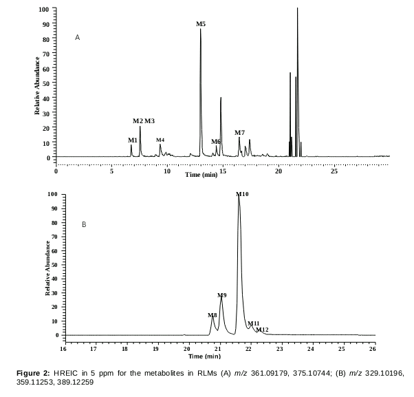
The high–resolution extracted Ion Chromato-graphy (HREIC) of blank, GA incubated in RLMS samples in 30 min are shown in the . For the first time, a total of 12 metabolites of GA were detected and identified based on accurate mass measurements, fragmentation patterns, and chromatographic retention times in positive ion mode. The detailed information is illustrated in .
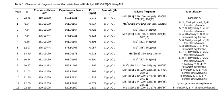
Metabolite M0, M8 and M12 were identified as GA, 5-hydroxy-7, 3', 4', 5'-tetramethoxyflavone and 5-hydroxy-7, 3', 4'-trimethoxyflavone, respectively by comparing the retention time, [M+H]+ ion and MS2 spectra with authentic references.
Metabolites M1, M2, M4, M6 and M7 were detected at 6.74, 7.54, 9.36, 14.40 and 16.44 min with protonated molecular ions [M+H]+ at m/z 361.09153 (-0.72 ppm, C18H17O8), m/z 361.09164 (-0.43 ppm, C18H17O8), m/z 361.09157 (-0.61 ppm, C18H17O8), m/z 361.09171 (-0.22 ppm, C18H17O8), and m/z 361.09168 (-0.30 ppm, C18H17O8), 58 Da (2CH2+OCH2) less than that of GA. The diagnostic product ions at m/z 346 [M+H- CH3。]+, m/z 314 [M+H- 3CH3。]+, and m/z 329[M+H- 2CH3。]+ were detected. They were tentatively identified as 8, 3’, 5’-trihydroxy-5, 7, 4’-trimethoxyflavone, 5, 7, 3’-trihydroxy-8, 4’, 5’-trimethoxyflavone, 5, 8, 3’-trihydroxy-7, 4’, 5’-trimethoxyflavone, 5, 7, 4’-trihydroxy-8, 3’, 5’-trimethoxyflavone, and 5, 8, 4’-trihydroxy-7, 3’, 5’-trimethoxyflavone, respectively [16].
Metabolites M3 and M5 were detected at 7.54 and 12.97 min respectively, with the same quasi-molecular ion [M+H]+ at m/z 375.11 (C19H19O8) that is 44 Da (CH2+OCH2) less than the parent compound GA. The CID product-ion spectrum of m/z 375 displayed three major fragment ions at m/z 360 ([M+H-CH3•]+), m/z 314 ([M+H-2CH3•-OCH3•]+) and m/z 329 ([M+H-CH3•-OCH3•]+). It was identified as the de-methoxylation and de-methylation product of GA and were tentatively confirmed as 5, 4’-dihydroxy-7, 8, 3', 5'-tetra-methoxyflavone and 5, 3'-dihydroxy-7, 8, 4', 5'-tetramethoxyflavone, respectively [12].
Metabolite M9-M11 was eluted at 21.03, 21.60, and 22.00min, respectively. Each of them showed a protonated molecule ion at m/z 389.12 (C21H27O11), 30 Da (OCH2) more than that of parent drug. The fragment ions at m/z 374 (4.2 ppm, C6H9O7), m/z 359 (4.8 ppm, C6H9O7) and m/z 356 (4.8 ppm, C6H9O7) in the MS2 spectra of metabolite M9-M11, indicated that they were a pair of hydroxy-pentamethoxyflavone isomers. Based on examination of known polymethoxy-flavonoids isolated from genus Murraya, and from bibliography data [5,12], M9 and M10 were tentatively identified as 5-hydroxy-6, 7, 3', 4', 5'- pentamethoxyflavone, 5-hydroxy-6, 7, 8, 3', 4'-pentamethoxyflavone, respectively. M11 was identified as hydroxypentamethoxy-flavone.
Discussion
In the study, we identified metabolites of GA in vitro for the first time. These metabolites are polymethoxylated flavonoids with many methoxy groups (OH-PMFs), which facilitate their detection by ESI in positive mode. MS conditions were optimized on an UHPLC-ESI-LTQ-Orbitrap instrument using standard solution of GA (10 μg/mL). In order to obtain an appropriate elution system, two different mobile systems, acetonitrile – water and methanol – water were tested. It was found that for GA, [M+H]+ ions of basically the same intensity were commonly detected with high sensitivity with these two solvent conditions. However, the mobile systems of acetonitrile – water with a gradient elution mode afforded much lower column pressure and better resolution of chromatographic peaks among the metabolites and endogenous components than that of water–methanol.
To our best knowledge, not much has been done on the elucidation of the metabolites of GA, although some studies on the metabolism of GA have been done in rats [12]. For example, 26 metabolites of GA were unambiguously and tentatively identified in Sprague-Dawley rats by comparison of retention times and mass spectrometry. However, it is worth mentioning that in vitro metabolites of GA; and the differences between in vivo and in vitro metabolites of GA were still unclear until now. In our study, 12 metabolites were detected and identified in RLMs. There was a good agreement between these metabolites and those obtained in in vivo studies. This clearly shows that it is feasible to obtain metabolites of GA in vitro.
Conclusion
This study has successfully elucidated the in vitro metabolism of GA in RLMs using UHPLC-LTQ-Orbitrap mass spectrometry. Among the 12 metabolites detected, 3 have been unambiguously confirmed, while the others are tentatively identified. The structures clearly indicate that de-methoxylation and de-methylation are the major metabolic fates of GA. These metabolites of GA in RLMs would facilitate better understanding of the metabolism and its pharmacological effects.
Declarations
Acknowledgement
References
Archives


News Updates